
Page 228 - 73_04
P. 228
ENRIQUE J. DE LA ROSA Y COLS. ANAL. REAL ACAD. NAC. FARM.
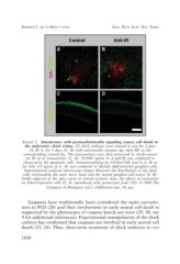
FIGURE 3. Interference with proinsulin/insulin signaling causes cell death in
the embryonic chick retina. E2 chick embryos were treated in ovo for 2 days
(A, B) or for 4 days (C, D) with anti-insulin receptor Igs (Anti-IR) or the
corresponding control Igs. The neuroretinas were then processed in whole-mount
(A, B) or as cryosections (C, D). TUNEL (green in A and B) was employed to
characterize the apoptotic cells. Immunostaining for G4/Ng-CAM (red in A, B) or
for Islet 1/2 (green in C, D) was employed to identify differentiated ganglion cells.
Superimposed confocal microscope images illustrate the distribution of the dead
cells surrounding the optic nerve head and the retinal ganglion cell axons (A, B).
Fields adjacent to the optic nerve in retinal sections show the effects of treatments
on Islet1/2-positive cells [C, D: reproduced with permission from (20); © 2000 The
Company of Biologists Ltd.]. Calibration bar, 50 µm.
Caspases have traditionally been considered the main executio-
ners in PCD (28) and their involvement in early neural cell death is
supported by the phenotypes of caspase knock-out mice (29, 30, see
9 for additional references). Experimental manipulation of the chick
embryo has confirmed that caspases are involved in early neural cell
death (23, 24). Thus, short-term treatment of chick embryos in ovo
1038