
Page 223 - 73_04
P. 223
VOL. 73 (4), 1031-1045, 2007 EARLY NEURAL CELL DEATH: AN OVERLOOKED...
neurons and glia, and it has been clearly recognized as a fundamental
element in generating and refining the complexity in the nervous
system. For example, differentiated neurons that do not succeed in
establishing the appropriate synaptic connections or that show
impaired electrical activity are selectively eliminated by PCD in order
to yield a highly specialised functional network. At this stage, the
death/survival decision in neurons is tightly regulated and depends
on the availability of neurotrophic factors as well as on cell-cell
interactions (1-6).
In striking contrast to the extensively studied cell death of mature
neurons, little attention has been paid to the PCD that affects
proliferating neuroepithelial cells and recently born neuroblasts (7-9).
For the last 12 years, we have studied this early phase of cell death in
the developing nervous system, characterizing the cell populations
affected, the regulatory mechanisms involved, its magnitude and the
functional implications of this process. In the present review, we shall
summarize all these aspects of early neural cell death, emphasizing
that our limited knowledge of this process may be hindering the
integrated understanding of neural development and vindicating the
more extensive study of this process.
CELL DEATH OCCURS DURING EARLY STAGES OF
NEURAL DEVELOPMENT
The nervous system mostly derives from the neural tube, a
structure generated from the embryonic ectoderm through inductive
interactions and morphogenic movements. Since the seminal work
of Glücksmann (10; see 7 for additional references), apoptotic cells
have been clearly identified during neural induction and neurulation
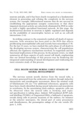
in vertebrates. In the neurulating chick embryo, apoptotic cells can
be observed when the neural tube is forming and regio-
nal specification is taking place. Indeed, TdT-mediated dUTP nick-
end labelling (TUNEL) of fragmented DNA, a hallmark of apoptosis,
reveals dying cells in precise locations of the embryo, for instance
at the anterior neuropore, the dorsal part of some prosomeres
and rhombomeres, the presumptive anlage of the otic vesicle (Fi-
gure 1, A-D).
1033