
Page 226 - 73_04
P. 226
ENRIQUE J. DE LA ROSA Y COLS. ANAL. REAL ACAD. NAC. FARM.
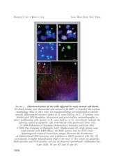
FIGURE 2. Characterization of the cells affected by early neural cell death.
E5 chick retinas were dissociated and stained with DAPI to visualize the nuclear
morphology (blue in A-C). Islet 1/2 immunostaining was performed to identify
recently differentiated neurons (green in D, same field as in C). E5 retinas were
labeled with [3H]-thymidine, dissociated and processed for autoradiography to
detect proliferating cells (grains in B, same field as in A). Arrowheads indicate the
pyknotic nuclei of apoptotic cells [reproduced with permission from (35];
© 1999 Federation of European Neuroscience Societies and from (20);
© 2000 The Company of Biologists Ltd.]. Whole-mount E5 chick retinas were
triple-stained with DAPI (blue), for BrdU (green) and for TUJ1 (red).
Superimposed confocal microscope images illustrate the distribution
of differentiated (TUJ1-positive) and proliferative (BrdU-positive) cells (E). (F)
corresponds to higher magnification field of the inset in (E), where an apoptotic,
BrdU-positive and TUJ1-positive cell can be observed (arrowhead). Calibration bar,
5 µm (A-D), 50 µm (E) and 10 µm (F).
1036