
Page 225 - 73_04
P. 225
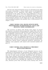
VOL. 73 (4), 1031-1045, 2007  EARLY NEURAL CELL DEATH: AN OVERLOOKED...

    We have also characterized the process of cell death in the chick
and mouse neuroretina, a classic model system in developmental
neurobiology (11-14). Apoptotic cells appear throughout the
development of the retina, from the early proliferative to the late
synaptogenic stages (15, 16; see 17 for additional references.). Even
at early developmental stages when neurogenesis begins, distinctive
and prominent patterns of cell death can be seen, following the
centro-peripheral gradient that parallels that of differentiation (18)
(Figure 1, E-F).

           EARLY NEURAL CELL DEATH AFFECTS BOTH
        PROLIFERATING NEUROEPITHELIAL CELLS AND

                     RECENTLY BORN NEUROBLASTS

    The presence of dying cells during early stages of neural
development, neurulation and neurogenesis, implies that cells other
than connecting neurons are affected since such connections are not
yet established. Unfortunately, these dead cells have not been
specifically identified in most studies of normal or manipulated
embryos (see 7 and 9 for additional references). In the embryonic
chick retina, we have identified dead cells as having recently exited
S-phase of the cell cycle, since they could incorporate labelled DNA-
precursors shortly before displaying apoptotic phenotype. In
addition, some apoptotic cells express early neuronal markers (Fi-
gure 2).

          EARLY NEURAL CELL DEATH IS A PRECISELY
                              REGULATED PROCESS

    The physiological relevance of any biological process can be
confirmed by determining the mechanisms that regulate it, which
also provides useful tools to define the magnitude and function, in
our case of early neural cell death. Interfering with cell death at
early stages produces abnormal development, as demonstrated by
embryonic and genetic manipulation. Knockout-mouse studies have
not only provided cues regarding the regulation of cell death but
also, dramatic proof that cell death occurs during early neural

                                                                                           1035