
Page 230 - 73_04
P. 230
ENRIQUE J. DE LA ROSA Y COLS. ANAL. REAL ACAD. NAC. FARM.
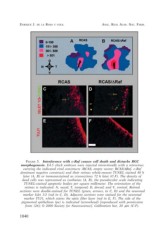
FIGURE 5. Interference with c-Raf causes cell death and disturbs RGC
morphogenesis. E4.5 chick embryos were injected intravitreally with a retrovirus
carrying the indicated viral constructs (RCAS, empty vector; RCAS/?Raf, c-Raf
dominant negative construct) and their retinas whole-mount TUNEL stained 48 h
later (A, B) or immunostained as cryosections 72 h later (C-F). The density of
dead cells was represented as isothanas (A, B), the pseudocolor scale indicating
TUNEL-stained apoptotic bodies per square millimeter. The orientation of the
retinas is indicated: N, nasal; T, temporal; D, dorsal; and V, ventral. Retinal
sections were double-stained for TUNEL (green, arrows, in C, D) and the neuronal
marker Islet 1/2 (red in C, D). Adjacent sections were stained for the neuronal
marker TUJ1, which stains the optic fiber layer (red in E, F). The side of the
pigmented epithelium (pe) is indicated (arrowhead) [reproduced with permission
from (26); © 2000 Society for Neuroscience]. Calibration bar, 20 µm (C-F).
1040