
Page 229 - 73_04
P. 229
VOL. 73 (4), 1031-1045, 2007 EARLY NEURAL CELL DEATH: AN OVERLOOKED...
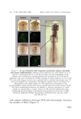
FIGURE 4. In ovo treatment with exogenous proinsulin reduces cell death
and causes malformations. Embryos were treated in ovo with vehicle (A, C, E
and G) or proinsulin (B, D, F and H) and after 8 h, the morphology of the
embryos was evaluated by counterstaining with neutral red (A, B, E and F).
Apoptotic cells were visualized by TUNEL staining of whole embryos (C, D, G and
H), and serial images (z-axis) were captured with a confocal microscope every 10
µm and compiled. For orientation, the different regions shown are boxed in a
photograph of the whole embryo (I). The upper box corresponds to the
prosencephalon (A-D) and the lower box to neural tube and rostral somites (E-H).
The main morphological features are labeled: n, anterior neuropore; ov, optic
vesicle; nt, neural tube; s, somite. Arrows in (B) and (F) indicate the main
morphological abnormalities [reproduced with permission from (22); © 2003
European Molecular Biology Association]. Calibration bar, 120 µm (A-H)
and 200 µm (I).
with caspase inhibitors decreases PCD and interestingly, increases
the number of RGCs (Figure 7).
1039