
Page 78 - 66_04
P. 78
ARMIN WOLF ANAL REAL ACAD. FARM.
shrinkage with invagination of nuclear membrane (large arrow), and vacuoles (v).
Inset: higher magnification of area from left cell showing mitochondria with intact
cristae and membranes (arrowhead). (D) Cluster of apoptotic bodies. A variety of
organelles is included in the different bodies, and one contains nuclear fragments
with condensed chromatin (large arrow). Bars: 1.0 µm.
In addition, induction of dilated ER and vacuoles was observed.
At 22 hours, all CsA-treated cultures displayed necrotic changes,
including swelling of all cytoplasmic compartments, swelling and
disappearance of mitochondrial cristae, disintegration of membranes, and
accumulation of large, dense granules in the mitochondrial matrix. The
necrotic changes were more pronounced in cultures treated with 50 µM
CsA (Fig. 6A). There was no significant difference in necrosis in control
preparations and CsA-treated preparations after 4 hours of incubation
(summary of data in Table 1).
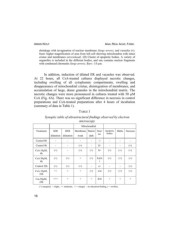
TABLE 1
Synoptic table of ultrastructural findings observed by electron
microscopy
Mitochondrial
Treatment SER RER Membrane Matriz Size/ Apoptotic Blebs Necrosis
no. bodies
dark
dilatation dilatation weak
Control 0h - - - - -/- - - -
Control 4h - - (+) - S/- - - (+)
CsA 10PM, (+) - (+) (+) S/- (+) (+) (+)
4h
CsA 50PM, (+) (+) + (+) S,d/n (+) (+) (+)
4h i
Control 22h (+) (+) (+) - s/- - - (+)
CsA 10PM, ++ + + (+) s/ni (+) (+) (+)
22h
Csa 50PM, +++ + + + d/ni + ++
22h
(+) marginal, + slight, ++ moderate, +++ marget, - no alteration/finding, s = swollen,
16