
Page 79 - 66_04
P. 79
VOL 66 (4) 2000 IN VITRO INDUCTION OF APOPTOSIS BY CYCLOSPORINE A
i = size increased, d = size decreased, ni = number increased
Biochemical markers of apoptosis
Four hours after CsA treatment, Annexin V reaction was increased
from 8 % in control preparations to 20 % in hepatocytes treated with CsA
(50 µM). The Annexin V reaction showed a similar
concentrationdependency as chromatin condensation/fragmentation and
DNA fragmentation. Twenty hours after CsA treatment, Annexin V-
stained cells increased from 7 % in controls to 30 % (Fig. 7).
After 20 hours of CsA incubation, the activity of the cysteine
protease caspase-3 and caspase-6, but not caspase-1, was statistically
significantly increased in comparison with controls. CsA treatment at 50
µM resulted in a sevenfold increase in caspase-3 activity; caspase-6
activity increased by 40 %. At the earlier time point (4 hours) no
statistically significant increases were seen (Fig. 8).
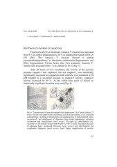
Fig. 6: Transmission electron micrograph of rat hepatocytes. (A) Control culture, 22
hours. Normal cell (upper third), and altered cell showing swelling of all cytoplasm
compartments, swelling of mitochondria and disappearance of mitochondrial cristae
(arrowhead), and detachment of ribosomes from rough endoplasmic reticulum
membranes and vacuolisation (small arrow). Glycogen (g), mitochondria (m),
vacuoles (v). (B) Culture treated with 50 µM CsA for 22 hours. Secondary necrosis
of apoptotic body: condensed chromatin (large arrows) and mitochondrial and
cytoplasmic fragments (small arrow). Inset: higher magnification of necrotic
17