
Page 52 - 81_02
P. 52
Andrea Aguado et al.
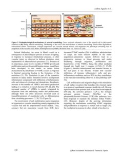
A B VSMC?hyperplasia
ARTERIAL?REMODELING
NORMAL Collagen
Endothelium deposition
Intima Collagen MMPs
Cytokines
VSMC Calcification Increased
Elastic Synthetic VSMC wall thickness
Fibers
Media
Osteogenic
VSMC
Adventitia
Pericytes/mesenchymal stem cells
Figure 3. Pathophysiological mechanisms of arterial remodeling. Cross sectional schematic view of the arterial wall in (A) normal
situation or (B) during arterial remodeling. Thickening of the wall is the main feature of arterial remodeling. Elastic fiber degradation,
extracellular matrix calcification, collagen deposition and vascular smooth muscle cell migration and phenotype switching lead to
adaptation of the vascular wall. Matrix metalloproteinase (MMP). Modified from van Varik et al. (20).
Intimal thickening can occur in blood vessels as a increased VSMC number (24). In addition, administration
consequence of physiological process as occurs in ageing, of AngII, the main effector peptide of the renin-
in response to increased intraluminal pressure, or after angiotensin-aldosterone system (RAAS) lead to a
vascular injury as observed in balloon dilatation, stent progressive increase in blood pressure and media
implantation or atherosclerosis processes (21). Because of thickening through migration, proliferation and
its importance, many in vivo models of VSMC growth and hypertrophy of the VSMCs, being this effect mediated
proliferation such as the carotid ligation mouse model have through the AngII type 1 receptor (AT1R) (7, 25-28)
been developed. In this model, an intima lesion (Figure 4). Besides hemodynamic and humoral factors, in
characterized by enrichment of VSMCs occurs in response the last years it has become evident that vascular
to luminal narrowing leading to the formation of the infiltration of immune inflammatory cells and pro-
neointima (19, 21). Neointima is part of the reparative inflammatory mediators such as ROS are key contributors
response to injury and its formation involves an important to the vascular remodeling observed in this pathology (29-
inflammatory component with infiltration of inflammatory 31).
cells and release of cytokines and chemokines, thrombosis,
increase in the number of VSMCs and matrix production Cell proliferation and migration begin with stimulation
leading to a reduction in vessel diameter (19, 22, 23). The of cell surface receptors that transduce the external signal
increased number of VSMCs is mainly originated by to a series of coordinated responses inside the cell. Diverse
migration from the underlying media and proliferation, signal transduction systems such as nuclear factor-kappa B
although there are other processes involved such as (NF-kB), the activator protein-1 (AP-1), the mitogen
transdifferentiation of endothelial cells or differentiation activated protein kinases (MAPKs) or the
from circulating precursors (7, 20, 21) (Figure 3). phosphatidylinositol-3-kinase (PI3K)/Akt pathways have
been proposed to translate the stimulus within VSMCs
The involvement of cell proliferation and/or migration (32). However, despite of the growing information
in hypertensive vascular remodeling mainly depends of the regarding the mechanisms controlling VSMC migration
vascular bed and the experimental model studied. Thus, and proliferation in response to stimuli such as AngII (28),
coronary but not mesenteric vessels from SHR show the regulation in response to other stimuli is less known.
132 @Real Academia Nacional de Farmacia. Spain