
Page 57 - 81_02
P. 57
Reactive oxygen species and vascular remodeling in cardiovascular diseases
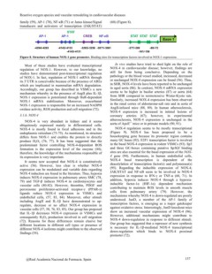
family (59), AP-1 (70), NF-?B (71) or Janus kinase/Signal (60) (Figure 8).
transducers and activators of transcription (JAK/STAT)
AP-1 STAT CREB NF-?B STAT STAT STAT Exon 1
AP-1
-4294/-4283 -4143/-4133 -3263/-3256 -3071/-3061 -277/-269 -80/-72
-4143/-4131 -161/-156
Figure 8. Structure of human NOX-1 gene promoter. Binding sites for transcription factors involved in NOX-1 expression.
Most of these studies have evaluated transcriptional In vivo studies have tried to shed light on the role of
regulation of NOX-1. However, to our knowledge, no NOX-4 in cardiovascular disease; however, findings are
studies have demonstrated post-transcriptional regulation still far from being conclusive. Depending on the
of NOX-1. In fact, regulation of NOX-1 mRNA through pathology or the blood vessel studied, increased, decreased
its 3’UTR is conceivable because of the presence of AREs or unchanged NOX-4 expression can be found (56). Thus,
which are implicated in mammalian mRNA degradation. in SHR, NOX-4 levels have been reported to be unchanged
Accordingly, our group has described in VSMCs a new in aged aorta (86). In contrast, NOX-4 mRNA expression
mechanism whereby in the presence of AngII plus IL-1ß, seems to be higher in basilar arteries (87) or aorta (64)
NOX-1 expression is potentiated through HuR-dependent from SHR compared to normotensive Wistar-Kyoto rats.
NOX-1 mRNA stabilization. Moreover, exacerbated Similarly, increased NOX-4 expression has been observed
NOX-1 expression is responsible for an increased NADPH in the renal cortex of aldosterone-salt rats and in aorta of
oxidase activity, ROS production and cell migration (72). AngII-infused mice (88, 89). In human atherosclerosis,
NOX-4 expression is increased in intimal lesions of
3.1.b. NOX-4 coronary arteries (67); however, in experimental
atherosclerosis, NOX-4 expression is unchanged in the
NOX-4 is very abundant in kidney and it seems aorta of ApoE-/- mice or in primate models (90, 91).
ubiquitously expressed mainly in differentiated cells.
NOX-4 is mostly found in focal adhesions and in the NOX-4 regulation seems to be mostly transcriptional
endoplasmic reticulum (73-75). As mentioned, its structure (Figure 9). NOX-4 has been proposed to be a
differs from NOX-1 and enables the protein to directly housekeeping gene because its promoter region contains
produce H2O2 (76, 77). It has been suggested that the many GC bases (92). E2F1 transcription factor is involved
predominant factor controlling NOX-4-dependent ROS in the basal NOX-4 expression in rodent VSMCs (93). Sp3
formation is the expression level of the enzyme (44); and three GC-boxes containing putative Sp/Klf binding
therefore, the knowledge of the mechanisms responsible of sites are also essential for the basal expression of the NOX-
its expression is very important. 4 gene (94). Furthermore, in human endothelial cells,
NOX-4 basal transcription is dependent of the
It seems now accepted that NOX-4 is constitutively deacetylation of transcription factor(s) and polymerase(s)
active (56). However, less clear is whether NOX-4 (95). Regarding the inducible expression of NOX-4,
expression can be modulated and variable data regarding JAK/STAT and NF-?B seem to be involved in NOX-4
NOX-4 induction are found in the literature. Thus, hypoxia expression in response to IFN-? or TNF-a (60, 71). In
induces NOX-4 expression in pulmonary artery SMC (78, addition, hypoxia induces NOX-4 through a hypoxia-
79) and TGF-ß induces NOX-4 in cardiomyocytes and inducible factor-1a (HIF-1a) dependent mechanism
vascular cells (80-82). However, thrombin, PDGF and contributing to maintain ROS levels in smooth muscle
peroxisome proliferator-activated receptor-? (PPAR-?) cells from pulmonary artery (79). However, the
ligands reduce NOX-4 expression in VSMCs and mechanisms whereby NOX-4 is down-regulated are poorly
endothelial cells, (57, 83, 84). Moreover, other stimuli understood. JunD, a member of the AP-1 family of
including AngII and IL-1ß have demonstrated to up- transcription factors, is emerging as a major gatekeeper
regulate, decrease or no affect NOX-4 expression in against oxidative stress. Interestingly, JunD knockout mice
vascular cells (57, 58, 76, 83, 85). Our group has proposed show an increased vascular expression of NOX-4 (96).
that IL-1ß decreases NOX-4 expression in VSMCs and However, additional mechanisms might contribute to
consequently H2O2 production involved in cell migration NOX-4 down-regulation in response to different stimuli.
(72). Reasons for these differences remain elusive but Our group has suggested that a repressor of new synthesis
different locations in different cell types or presence of is necessary for IL-1ß-mediated NOX-4 transcriptional
different NOX-4 isoforms might contribute to the observed down-regulation which binds to NOX-4 proximal
findings (54). promoter (72).
@Real Academia Nacional de Farmacia. Spain 137