
Page 50 - 81_02
P. 50
derived ROS released from phagocytic and vascular cells Andrea Aguado et al.
are involved in many processed associated to
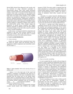
cardiovascular disease. Thus, many studies performed amounts of ECM. The tunica media is separated from the
mainly in animal models have demonstrated that increased tunica adventitia by a second layer of elastic fibers, the
oxidative stress state is necessary for the initiation and external elastic lamina. In response to different vasoactive
progression of vascular disease that may ultimately lead to factors and hemodynamic forces, VSMCs can release a
heart attack and strokes. Some members of the NADPH variety of substances which affect vascular tone and
oxidases are constitutively expressed in the vasculature. structure.
However, different hormones, inflammatory mediators or
hemodynamic stimuli important in cardiovascular diseases, - Adventitia: it is mainly formed by fibroblasts but it
increase the activity or the expression of NADPH oxidase also contains macrophages and mast cells and different
isoforms leading to a deleterious oxidative stress status in components of the ECM. In the last years, it has become
the cardiovascular system. These changes trigger the evident that the adventitia is not only a mechanical support
production of growth factors, proteases and cellular for the vessel but also an active player of the regulation of
adhesion molecules by different vessel cell types leading vascular tone and structure by releasing different factors.
to structural changes in the wall of the vessel in a process
known as vascular remodeling. The ECM is a gel-like form which functions as a
scaffolding structure for the vascular cells and determines
In this review, we discuss in detail the composition and the elasticity and mechanical properties of the vessels.
regulation of the main NOX enzymes expressed in the Their components are synthetized by different cell types of
media layer (NOX-1 and 4) and their roles in vascular the vascular wall. The two main ECM proteins are
remodeling associated with cardiovascular diseases. collagen and elastin; while elastin confers the elastic
2. VASCULAR REMODELING properties to vessels, collagen provides the strength (4).
2.1. Artery structure There are other ECM proteins that are in less quantity such
as glycoproteins, proteoglycans and integrins that are
Arteries are divided in three concentrical layers from involved in several cellular processes (4). Among them,
the inside out: intima, media and adventitia which are tenascin-C (TN-C), which is an inducible glycoprotein,
organized in cellular components and extracellular matrix expressed predominantly in embryonic, remodeled adult
(ECM) (Figure 1). tissues and in pathological conditions, is particularly
interesting. Competitive binding of TN-C to ECM proteins
Figure 1. Artery structure. Wall section showing all layers of and their counterpart cell-surface receptors mediates its
an artery wall. ability to modulate cell-ECM interactions. The capacity of
TN-C to interact with a wide range of ECM molecules
- Intima: it is in the inner part of the vessel and may also enable it to contribute to the structural
comprises a monolayer of endothelial cells which lay in organization of the ECM. In addition, TN-C can promote
the basement membrane. This layer of endothelial cells is migration and proliferation by direct activation of cell-
separated from the media layer by the internal elastic surface growth factor receptors and cellular differentiation
lamina which is a fenestrated lamina of elastic fibers. The by up-regulating androgen receptor and endothelin type 1
intima layer is important in the control of vascular function receptor expression (5). Thus, TN-C relevance relies on its
and structure because endothelial cells are an important implication in vascular cell differentiation, proliferation
source of vasoconstrictor/vasodilator and and migration (5).
proliferative/antiproliferative factors.
2.2. Types of vascular remodeling
- Media: this layer includes circumferentially arranged
vascular smooth muscle cells (VSMCs) and variable It is now accepted that the vascular wall can change its
structure in order to maintain the appropriate lumen size to
130 permit normal blood flow. This process is termed vascular
remodeling (6). This ability of the arteries to adapt its
structure in response to physiological and pathological
conditions is essential in situations such as pregnancy or
aging but also in many arterial diseases. Thus, the inability
of the vessels to remodel appropriately is considered a
form of “vascular failure” that can lead to pathologic states
such as hypertension, atherosclerosis or restenosis (7).
This process is active and involves structural changes
including cell growth, death, migration and the synthesis
or degradation of the ECM (7).
Vascular remodeling can occur with or without growth
of the vessel wall (i.e. hypertrophic, eutrophic or
hypotrophic) and with smaller, greater or similar lumen
size (inward, outward or compensated) (8). Vascular
remodeling often differs depending on the vessel type or
the cardiovascular disease model (Figure 2).
@Real Academia Nacional de Farmacia. Spain