
Page 117 - 80_02
P. 117
The
role
of
the
catecholaminergic…
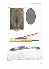
Figure
4.
Techniques
to
manipulate
gene
expression
in
the
chick
embryo:
implanted
microbeads
and
electroporation.
A)
The
stage
3--5
blastoderm
can
be
accessed
in
ovo
and
B)
a
factor--coated
microbead
(yellow
circle)
can
be
implanted
at
the
desired
location
lateral
to
the
heart
field
of
one
side
(blue
square).
After
6--18
h
of
further
incubation
the
embryo
can
be
retrieved
and
processed
for
RNA
or
protein
detection.
C)
The
blastoderm
can
also
be
placed
in
culture
and
injected
and
electroporated
with
DNA
constructs,
such
as
that
shown
in
schematic
form,
in
which
the
sequences
of
TH
and
the
green
fluorescence
protein
(GFP)
are
linked
by
an
IRES
(Internal
Ribosome
Entry
Site).
After
18--36
h
of
additional
incubation,
embryos
can
be
collected
and
processed.
353
