
Page 83 - 83_01
P. 83
Targeting inflammation in the retina: a new therapeutic approach in diabetic retinopathy
!
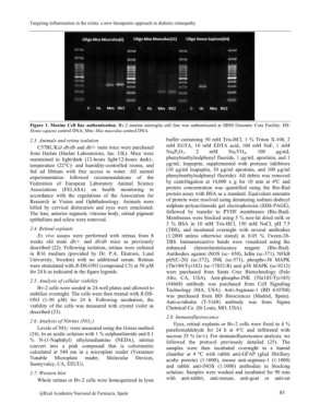
Figure 1. Murine Cell line authentication. Bv.2 murine microglia cell line was authenticated at IIBM Genomic Core Facility. HS:
Homo sapiens control DNA; Mm: Mus musculus control DNA.
2.3. Animals and retina isolation buffer containing 50 mM Tris-HCl, 1 % Triton X-100, 2
mM EGTA, 10 mM EDTA acid, 100 mM NaF, 1 mM
C57BL/KsJ db/db and db/+ male mice were purchased Na4P2O7, 2 mM Na3VO4, 100 µg/mL
from Harlan (Harlan Laboratories, Inc. UK). Mice were phenylmethylsulphonyl fluoride, 1 µg/mL aprotinin, and 1
maintained in light/dark (12-hours light/12-hours dark)-, µg/mL leupeptin, supplemented with protease inhibitors
temperature (22°C)- and humidity-controlled rooms, and (10 µg/ml leupeptin, 10 µg/ml aprotinin, and 100 µg/ml
fed ad libitum with free access to water. All animal phenylmethylsulphonyl fluoride). All debris was removed
experimentation followed recommendations of the by centrifugation at 14,000 x g for 10 min at 4ºC and
Federation of European Laboratory Animal Science protein concentration was quantified using the Bio-Rad
Associations (FELASA) on health monitoring in protein assay with BSA as a standard. Equivalent amounts
accordance with the regulations of the Association for of protein were resolved using denaturing sodium dodecyl
Research in Vision and Ophthalmology. Animals were sulphate-polyacrilamide gel electrophoresis (SDS-PAGE),
killed by cervical dislocation and eyes were enucleated. followed by transfer to PVDF membranes (Bio-Rad).
The lens, anterior segment, vitreous body, retinal pigment Membranes were blocked using 5 % non-fat dried milk or
epithelium and sclera were removed. 3 % BSA in 10 mM Tris-HCl, 150 mM NaCl, pH 7.5
(TBS), and incubated overnight with several antibodies
2.4. Retinal explants (1:2000 unless otherwise stated) in 0.05 % Tween-20-
TBS. Immunoreactive bands were visualized using the
Ex vivo assays were performed with retinas from 8 enhanced chemioluminiscence reagent (Bio-Rad).
weeks old male db/+ and db/db mice as previously Antibodies against iNOS (sc- 650), I?Ba (sc-371), NFkB
described (22). Following isolation, retinas were cultured p65(C-20) (sc-372), JNK (sc-571), phospho-38 MAPK
in R16 medium (provided by Dr. P.A. Ekstrom, Lund (Thr180/Tyr182) (sc-17852-R) and p38 MAPK (sc-9212)
University, Sweden) with no additional serum. Retinas were purchased from Santa Cruz Biotechnology (Palo
were stimulated with R-DS-ONJ (compound C5) at 50 µM Alto, CA, USA). Anti-phospho-JNK (Thr183/Tyr185)
for 24 h as indicated in the figure legends. (#4668) antibody was purchased from Cell Signaling
Technology (MA, USA). Anti-Arginase-1 (BD 610708)
2.5. Analysis of cellular viability was purchased from BD Biosciences (Madrid, Spain).
Anti-a-tubulin (T-5168) antibody was from Sigma
Bv-2 cells were seeded in 24-well plates and allowed to Chemical Co. (St Louis, MO, USA).
stabilize overnight. The cells were then treated with R-DS-
ONJ (1-50 µM) for 24 h. Following incubation, the 2.8. Immunofluorescence
viability of the cells was measured with crystal violet as
described (23). Eyes, retinal explants or Bv-2 cells were fixed in 4 %
paraformaldehyde for 24 h at 4°C and infiltrated with
2.6. Analysis of Nitrites (NO2-) sucrose 25 % (w/v). For immunofluorescence analysis, we
Levels of NO2- were measured using the Griess method followed the protocol previously detailed (25). The
samples were then incubated overnight in a humid
(24). In an acidic solution with 1 % sulphanilamide and 0.1 chamber at 4 ºC with rabbit anti-GFAP (glial fibrillary
% N-(1-Naphthyl) ethylenediamine (NEDA), nitrites acidic protein) (1:1000), mouse anti-arginase-1 (1:1000)
convert into a pink compound that is colorimetric and rabbit anti-iNOS (1:1000) antibodies in blocking
calculated at 540 nm in a microplate reader (Versamax solution. Samples were washed and incubated for 90 min
Tunable Microplate reader, Molecular Devices, with anti-rabbit, anti-mouse, anti-goat or anti-rat
Sunnyvaley, CA, EEUU).
2.7. Western blot
Whole retinas or Bv-2 cells were homogenized in lysis
@Real Academia Nacional de Farmacia. Spain 83