
Page 126 - 82_02
P. 126
acute liver injury María Ángeles Pajares
The results described in the previous section suggested correlating with a severe alteration in the isoenzyme
that changes in MATa1 subcellular distribution might be pattern according to AGFC profiles, where only a small
related to disease development. In order to examine this amount of MAT III was detected (93)(Figure 5).
possibility, we choose two rat models of acute liver injury Altogether these results followed the previously published
that were previously used for partial characterization of pattern detected by activity measurements in the cytosol
alterations in cytosolic methionine metabolism (89-91). (89). Paracetamol intoxication produced a more modest
Precisely, D-galactosamine and paracetamol intoxications decrease in cytosolic MATa1 protein levels together with
were known to induce reductions in MAT activity and a completely different pattern in the MAT isoenzyme
SAM levels (89,90,92). These changes in the D- profile by AGFC, where an increase in MAT I content was
galactosamine model were ascribed to an anomalous ratio readily observed. Moreover, while the MAT III/I activity
of the MAT isoenzyme activities towards enhanced MAT ratio increased in the cytosol of paracetamol treated livers,
III activity (89,90). We were able to reproduce these the opposite trend was followed by the protein ratio, thus
previous results in the cytosol, reductions in total MAT suggesting a certain degree of inactivation of MAT I in
activity being more modest for paracetamol than for D- these samples (93).
galactosamine treatments (92,93). The mechanism of
action of both drugs is quite different, D-galactosamine Analysis of the nuclear effects of the treatments
inducing depletion of the uridine pool (94), and thus revealed accumulation of MATa1 in this compartment
highlighting the interest of analyzing putative expression both by D-galactosamine and paracetamol intoxications
changes in that model. After 48 hours of D-galactosamine (93). AGFC revealed increased levels of MAT I and a
treatment, changes in the mRNA levels of most genes in reduction in those of MATa1 monomers in nuclear
the methionine cycle were evident, namely: i) the fractions of D-galactosamine-treated livers, which
Mat1a/Mat2a expression switch, followed by a more correlated with the detection of enhanced nuclear MAT
modest increase in Mat2b expression; ii) increased mRNA activity (Figure 5). In contrast, paracetamol induced a
levels for Ahcy and Mtr; iii) enhanced expression of genes modest increase in nuclear MAT I levels, but no
involved in glutathione synthesis; and iv) decreased significant changes in activity were measured. However, in
expression of Gnmt and Bhmt (93). In general, genes both models enhanced levels of the me3K27H3 repression
considered "exclusively hepatic" showed lower expression. mark were also detected. This lack of correlation between
activity and the histone modification in paracetamol
Additional analyses carried out showed that the intoxication prompted us to evaluate nuclear SAM levels.
changes induced by D-galactosamine in the cytosolic However, the procedures needed to purify nuclear
fractions precluded this measurement, given the exchange
protein levels of MATa1, BHMT, SAHH and GNMT through the nuclear pore that takes place during the
closely matched the alterations observed in their extensive washing steps required for isolation. These
measurements will be only possible when appropriate
expression (93). Focusing specifically on MATa1, the imaging techniques become available.
protein levels were reduced by ~50%, this decrease
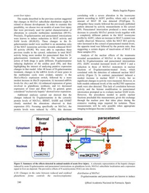
Figure 5. Summary of the effects detected in animal models of acute liver injury. A schematic representation of the hepatic changes
induced by acute D-galactosamine and paracetamol intoxications on glutathione levels, MATa1 subcellular localization and a repression
signal in liver are depicted. The preventive effects of N-acetylcysteine (NAC) are also shown.
4.10. Changes in the ratio between reduced and oxidized distribution of MATa1.
glutathione forms controls the nucleocytoplasmic D-galactosamine and paracetamol are known to induce
240 @Real Academia Nacional de Farmacia. Spain