
Page 121 - 82_02
P. 121
The new levels of redox regulation of S-adenosylmethionine synthesis
knowledge, several questions remained unanswered, being sample precluded crystallization. Next, we tried to use the
their response the objective of our group in the last 20 recombinant protein overexpressed in E. coli (53,54), and
years. several important problems had to be faced: 1) the level of
soluble protein was very low; 2) it was impossible to
4. OUR CONTRIBUTION TO UNDERSTAND separate the recombinant protein from the bacterial MAT
due to their high homology and identical chromatographic
MAMMALIAN METHIONINE behavior; and 3) refolding of MAT I/III from the inclusion
bodies required a protocol that had to be established.
ADENOSYLTRANSFERASES: FROM STRUCTURE Nevertheless, we decided to pursuit the design of a specific
refolding protocol (57), rendering large amounts of soluble
TO NEW REGULATORY LEVELS protein, and that latter revealed its use for additional
purposes. The success of this refolding protocol relied on
The objective since we started our independent work the use of very low protein concentrations, the addition of
by 1994 was to understand several aspects of MAT Mg2+ to the buffers, the utilization of two refolding steps
(fast and slow), and the maintenance of the 10 cysteines of
regulation that were poorly addressed, despite the intensive
work of several groups since their discovery. Some of the rat MATa1 in a reduced state during the whole procedure
(57). Characterization of the refolded protein showed that
limiting facts for the field at that time related to the both MAT I and MAT III isoenzymes could be obtained,
and their interconversion was possible just by
difficult purification procedures to obtain the isoenzymes concentration or dilution of the sample (57). Moreover,
kinetic parameters and circular dichroism spectra of the
and the lack of good antibodies or structural information. refolded proteins were similar to those of the isoenzymes
Therefore, our initial work was focused on these aspects to purified from rat liver (57,58). These facts, together with
the large amount of refolded and purified MAT I/III
develop better protocols and tools that allowed progress in obtained, led us to use these proteins in new crystallization
the study of MATs, and which latter favored additional attempts that were successful.
studies on animal models, as will be explained below.
4.1. Association of a-subunits is required to obtain active
MATs
The first objective of our work was to understand the
need for oligomerization of MAT a-subunits, and for this
purpose we undertook the crystallization of rat MAT I and
MAT III. Our initial attempts were based in the use of the
rat liver purified proteins, but the heterogeneity of the
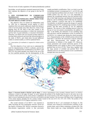
Figure 3. Structural details of MATa1 and its dimer. A cartoon representation of the secondary elements found in rat MATa1
monomers is shown in the upper left panel. An arrow indicates the position of the flexible loop of access to the active site in the
completely folded subunit and the central domain is highlighted. The crystal structure of the MAT III appears on the right side of the
figure, an arrow indicating the entrance to the active site located at the dimer interface. The lower part of the figure shows a magnification
of the central domain, including the five cysteine residues located in this area (C35, C61, C57, C67, C105). The position of the
intrasubunit disulfide (C35-C61) appears indicated by a circle.
The crystal structure of rat MAT I was reported in described for the E. coli counterpart (5) (Figure 3). The
2000, becoming the first mammalian structure solved for domains were formed by nonconsecutive stretches of the
this family of proteins (59). MATa1 monomers showed a protein chain, each of them containing a ß-sheet (59). The
three-domain organization similar to that previously three sheets made up a large hydrophobic surface that
@Real Academia Nacional de Farmacia. Spain 235