
Page 76 - 76_03
P. 76
ALBERTO BARTOLOMÉ Y COLS. AN. R. ACAD. NAC. FARM.
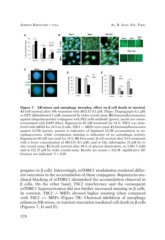
Figure 7. ER-stress and autophagy interplay, effect on ß cell death or survival.
A) Cell survival after 24h treatment with MG132 0,5 µM; Thaps: Thapsigargin 0,1 µM;
or DTT: dithiothreitol 5 mM, measured by violet crystal assay. B) Immunofluoresecence
against ubiquitin-protein conjugates with FK2 mAb antibody (green), nuclei are conun-
tersestained with DAPI (blue). Rapamycin 40 nM treatment for 24 h. TSC2 was inter-
fered with siRNA for 24 h in ß cells, TSC2 -/- MEFs were used. C) Immunofluorescence
against LC3B (green), puncta is indicative of lipidated LC3B accumulation in au-
tophagosomes, while cytoplasmic staining is indicative of no autophagic activity.
Rapamycin 40 nM was used for 24 h. D) Pancreatic ß-cell survival after 24 h treatment
with a lower concentration of MG132 (0,1 µM), and or CQ; chloroquine 25 µM by vi-
olet crystal assay. E) ß-cell survival after 48 h of glucose deprivation, or 2-DG 5 mM;
and or CQ 25 µM by violet crystal assay. Results are means ± S.E.M. significative dif-
ferences are indicated *P < 0.05.
gregates in ß cells. Interestingly, mTORC1 modulation rendered differ-
ent outcomes in the accumulation of these conjugates. Rapamycin-me-
diated blocking of mTORC1 diminished the accumulation observed in
ß cells. On the other hand, TSC2 interference and the consequent
mTORC1 hyperactivation did not further increased staining in ß cells.
In contrast, TSC2 -/- MEFs showed higher staining when compared
with TSC2 +/+ MEFs (Figure 7B). Chemical inhibition of autophagy
enhances ER-stress, or nutrient starvation mediated cell death in ß cells
(Figures 7, D and E).
370