
Page 72 - 76_03
P. 72
ALBERTO BARTOLOMÉ Y COLS. AN. R. ACAD. NAC. FARM.
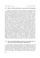
3.5. Effect of TSC2 interference on pancreatic ß cell signaling
To assess the importance of TSC complex in the integration of ß
cell signaling, we targeted the expression of TSC2 with siRNA. A sim-
ilar rate of TSC2 knockdown was achieved in all the cell lines, close
to 70% of protein expression interference (Figure 4B). TSC2 knock-
down induced p70S6K basal phosphorylation in all cell lines studied
(Figure 4A). TSC2 interference generated insulin resistance, decreas-
ing Akt and p70S6K phosphorylation in response to insulin in IR +/+
(Figure 4, A and C). However, this insulin resistance effect was de-
pendent on the IR isoform expression. Thus, Rec A, but not Rec B
cells overcome insulin resistance on Akt Ser473 and p70S6K phospho-
rylation (Figure 4, A, C and D). In addition, TSC2 interference gener-
ated insulin resistance, decreasing ERK 1/2 phosphorylation in IR +/+,
Rec A, or Rec B cell lines (Figure 4A). The insulin resistance induced
by TSC inactivation in IR +/+ or Rec B cells was blunted by rapamycin
treatment, indicating a requirement for mTORC1 signaling. In con-
trast, the effect observed on ERK 1/2 was rapamycin-independent in
all cell lines studied (Figure 4A). In IR -/- cells, TSC2 interference im-
paired glucose mediated ERK 1/2 phosphorylation. However, glucose
induced p70S6K signaling regardless of the presence of the MEK 1/2
inhibitor U0126 through ERK 1/2 independent pathway (Figure 4A,
upper right panel). To further investigate the differences between IRA
or IRB expression on insulin resistance, we stimulated cells with in-
sulin 10 nM for 15 minutes, and observed a different modulation of
IRS-1 Ser307 phosphorylation. This phosphorylation is mediated by
p70S6K, as described before (15), reverted by rapamycin, and upreg-
ulated in Rec B cells compared with Rec A or IR +/+ cells (Figure 5).
3.6. Role of the TSC2/mTORC1 pathway in the proliferation of
pancreatic ß cells bearing IRA or IRB isoforms
To assess the role played by mTORC1 in the proliferation rate of ß
cell lines studied, we submitted those cells to 10% FBS in the presence
or absence of rapamycin 40 nM (Figure 6). The rate of proliferation, as
estimated by cell counting, increased by 8-fold in IR +/+ or in Rec B
and by 4-fold in IR -/- cells (Figure 6, A and B). In addition, Rec A cells
showed a much higher rate of proliferation under the same experimen-
366