
Page 89 - 82_02
P. 89
Virginia Pardo Marqués, Águeda González-Rodríguez, Ángela Martínez Valverde
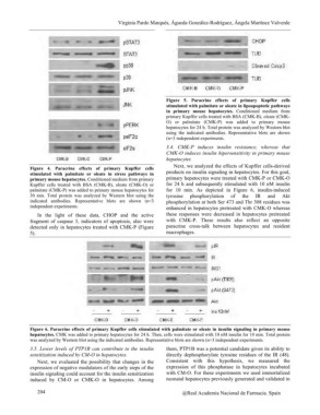
Figure 4. Paracrine effects of primary Kupffer cells Figure 5. Paracrine effects of primary Kupffer cells
stimulated with palmitate or oleate in stress pathways in stimulated with palmitate or oleate in lipoapoptotic pathways
primary mouse hepatocytes. Conditioned medium from primary in primary mouse hepatocytes. Conditioned medium from
Kupffer cells treated with BSA (CMK-B), oleate (CMK-O) or primary Kupffer cells treated with BSA (CMK-B), oleate (CMK-
palmitate (CMK-P) was added to primary mouse hepatocytes for O) or palmitate (CMK-P) was added to primary mouse
30 min. Total protein was analyzed by Western blot using the hepatocytes for 24 h. Total protein was analyzed by Western blot
indicated antibodies. Representative blots are shown (n=3 using the indicated antibodies. Representative blots are shown
independent experiments. (n=3 independent experiments.
In the light of these data, CHOP and the active 3.4. CMK-P induces insulin resistance, whereas that
fragment of caspase 3, indicators of apoptosis, also were CMK-O induces insulin hipersensitivity in primary mouse
detected only in hepatocytes treated with CMK-P (Figure hepatocytes
5).
Next, we analyzed the effects of Kupffer cells-derived
products on insulin signaling in hepatocytes. For this goal,
primary hepatocytes were treated with CMK-P or CMK-O
for 24 h and subsequently stimulated with 10 nM insulin
for 10 min. As depicted in Figure 6, insulin-induced
tyrosine phosphorylation of the IR and Akt
phosphorylation at both Ser 473 and Thr 308 residues was
enhanced in hepatocytes pretreated with CMK-O whereas
these responses were decreased in hepatocytes pretreated
with CMK-P. These results also reflect an opposite
paracrine cross-talk between hepatocytes and resident
macrophages.
Figure 6. Paracrine effects of primary Kupffer cells stimulated with palmitate or oleate in insulin signaling in primary mouse
hepatocytes. CMK was added to primary hepatocytes for 24 h. Then, cells were stimulated with 10 nM insulin for 10 min. Total protein
was analyzed by Western blot using the indicated antibodies. Representative blots are shown (n=3 independent experiments.
3.5. Lower levels of PTP1B can contribute to the insulin them, PTP1B was a potential candidate given its ability to
sensitization induced by CM-O in hepatocytes. directly dephosphorylate tyrosine residues of the IR (48).
Consistent with this hypothesis, we measured the
Next, we evaluated the possibility that changes in the expression of this phosphatase in hepatocytes incubated
expression of negative modulators of the early steps of the with CM-O. For these experiments we used immortalized
insulin signaling could account for the insulin sensitization neonatal hepatocytes previously generated and validated in
induced by CM-O or CMK-O in hepatocytes. Among
204 @Real Academia Nacional de Farmacia. Spain