
Page 43 - 81_03
P. 43
Functional characterization of P2Y1 and P2X4 receptors in human neuroblastoma SK-N-MC cells
P2Y6, P2Y11 and P2Y13 proteins can be detected in the most commonly utilized P2 agonists by single-cell
western blot assays (results not shown). Again, imaging after loading cells with the fluorescent calcium
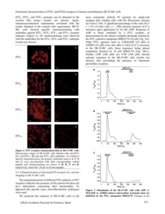
immunocytochemical experiments correlated with the dye Fura-2 AM. A significant percentage of the cells (91.5
results obtained in the western blot experiments: SK-N- ± 1.5% of total cells, n = 240) showed transient [Ca2+]i
MC cells showed specific inmmunostaining with increases upon application of 10 µM ADP. Response to
antibodies against P2Y1, P2Y6, P2Y11 and P2Y13 receptor ADP is likely mediated by a P2Y1 receptor, as
subtypes (Figure 2). No immunostaining were observed demonstrated by the almost complete blockade exerted by
with the antibodies for the P2Y2, P2Y4 and P2Y12 subtypes the P2Y1-selective antagonist MRS2179 (10 µM, Fig. 3A).
(results not shown). Other P2Y1 agonists such as 2-MeSADP (10 µM) or
ADPßS (10 µM) were also able to elicit [Ca2+]i increases
AB in the SK-N-MC cells, these responses being almost
completely blocked by 10 µM MRS2179 (Fig. 3B,C).
P2Y1 Neither UDP (100 µM) nor UTP (100 µM) elicited
calcium responses in the SK-N-MC cells (results not
shown), thus precluding the presence of functional
pyrimidine receptors.
CD
P2Y6
EF
P2Y11
GH
P2Y13
Figure 2. P2Y receptors immunodetection in SK-N-MC cells. Figure 3. Stimulation of the SK-N-MC cells with ADP, 2-
Fluorescence image of SK-N-MC cells labeled with anti-P2Y6 MeSADP or ADPßS induces intracelular transients that are
(C), anti-P2Y11 (E) and anti-P2Y13 (G) antibodies. To confirm a inhibited by the P2Y1 antagonist MRS2179. Changes in the
specific immunoreaction, the primary antibodies used in A, C, E
and G were pre-absorbed with their corresponding control 251
peptide and immunostaining are shown in B, D, F and H,
respectively. Scale bar= 20 µm in all micrographs.
3.3. Characterization of functional P2 receptors by calcium
imaging in SK-N-MC cells
The immunodetection of different P2X subunits or P2Y
receptors indicates the presence of the protein but does not
give information concerning their functionality. To
approach this specific issue, microfluorimetric techniques
were used.
We analyzed the response of SK-N-MC cells to the
@Real Academia Nacional de Farmacia. Spain