
Page 108 - 76_01
P. 108
ÓSCAR ESCRIBANO Y COLS. AN. R. ACAD. NAC. FARM.
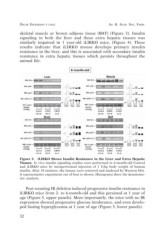
skeletal muscle or brown adipose tissue (BAT) (Figure 3). Insulin
signaling in both the liver and these extra hepatic tissues was
similarly impaired in 1 year-old iLIRKO mice, (Figure 4). These
results indicate that iLIRKO mouse develops primary insulin
resistance in the liver, and this is associated with secondary insulin
resistance in extra hepatic tissues which persists throughout the
animal life.
Figure 3. iLIRKO Shows Insulin Resistance in the Liver and Extra Hepatic
Tissues. In vivo insulin signaling studies were performed in 6-month-old Control
and iLIRKO mice by intraperitoneal injection of 1 U/kg body weight of human
insulin. After 10 minutes, the tissues were removed and analyzed by Western blot.
A representative experiment out of four is shown. Histograms show the densitome-
tric analysis.
Post-weaning IR deletion induced progressive insulin resistance in
iLIRKO mice from 2- to 6-month-old and this persisted at 1 year of
age (Figure 5, upper panels). More importantly, the mice with no IR
expression showed progressive glucose intolerance, and even develo-
ped fasting hyperglycemia at 1 year of age (Figure 5, lower panels).
32