
Page 148 - 72_01
P. 148
M. TERESA GÓMEZ-CASARES Y COLS. AN. R. ACAD. NAC. FARM.
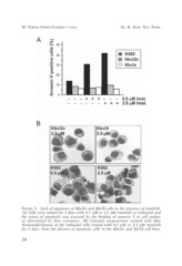
FIGURE 3. Lack of apoptosis of Kbcl2v and KbclX cells in the presence of imatinib.
(A) Cells were treated for 2 days with 0.5 µM or 2.5 µM imatinib as indicated and
the extent of apoptosis was assessed by the binding of annexin V to cell surface
as determined by flow cytometry. (B) Cytospin preparations stained with May
Grumwald-Giemsa of the indicated cells treated with 0.5 µM or 2.5 µM imatinib
for 2 days. Note the absence of apoptotic cells in the Kbcl2v and KbclX cell lines.
34