
Page 45 - 75_04
P. 45
VOL. 75 (4), 901-910, 2009 OVER-EXPRESSION OF P2Y2 RECEPTOR AFTER SILENCING...
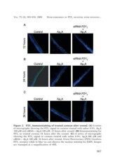
Figure 2. P2Y2 immunostaining of treated corneas after wound. (A) A series
of micrographs showing the P2Y2 signal in corneas treated with saline 0.9%, Ap4A
100 µM and siRNA + Ap4A 100 µM, 12 hours after wound. (B) Immunostaining for
P2Y2 in treated corneas 24 hours after the wound. (C) A series of micrographs
showing the P2Y2 signal in corneas treated with saline 0.9%, Ap4A 100 µM and
siRNA + Ap4A 100 µM, 36 hours after wound. Green fluorescence (FITC) localizes
P2Y2 receptor while in blue we can observe the nuclear staining for DAPI. Images
are managed at a magnification of 40X.
907