
Page 35 - 80_04
P. 35
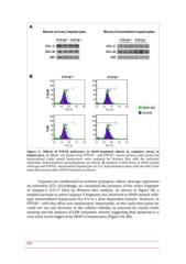
Figure 2.- Effects of PTP1B deficiency in APAP-mediated effects in oxidative stress in
hepatocytes. A. Whole cell lysates from PTP1B+/+ and PTP1B-/- mouse primary (left panel) and
immortalized (right panel) hepatocytes were analyzed by Western blot with the indicated
antibodies. Representative autoradiograms are shown. B. Analysis of ROS levels in APAP-treated
wild-type and PTP1B-/- immortalized hepatocytes for 6 h. Representative plots with the shift of the
mean fluorescence after APAP treatment are shown.

        Caspases are synthesized as inactive zymogens, whose cleavage represents
its activation (37). Accordingly, we examined the presence of the active fragment
of caspase-3 (15-17 kDa) by Western blot analysis. As shown in Figure 3B, a
marked increase in active caspase-3 fragment was observed in APAP-treated wild-
type immortalized hepatocytes for 8 h in a dose-dependent manner. However, in
PTP1B-/- cells this effect was ameliorated. Importantly, at this early time point we
could not see any decrease in the cellular viability as assessed by crystal violet
staining and the analysis of LDH enzymatic activity suggesting that apoptosis is a
very early event triggered by APAP in hepatocytes (Figure 4A, 4B).

674