
Page 86 - 76_01
P. 86
DAVID LEÓN NAVARRO Y COLS. AN. R. ACAD. NAC. FARM.
Immunocytochemical studies also demonstrate the presence
of ionotropic (such as P2X7, Figure 2A) and metabotropic (such
as P2Y1, Figure 2D) purinergic receptors in rat cultured cerebellar
granule neurons. However, these cells showed different calcium
responses to ATP. To determine the dose-response relations, neurons
were challenged with several concentrations of ATP. Results,
expressed as a percentage of cells responding per total number of
cells tested for each concentration, have shown the following values:
a) 1 µM ATP, 10%; b) 100 µM ATP, 30%; c) 200 µM ATP, 50% (not
shown). It is necessary to consider that we are only measuring
responses to added nucleotides, not to the hydrolysis products
(mainly adenosine) that could appear due to ectonucleotidases
action. The continuous superfusion of the cell chamber avoids
concentration of any hydrolytic product in the extracellular medium.
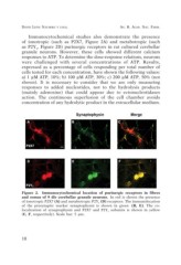
Figure 2. Immunocytochemical location of purinergic receptors in fibres
and somas of 9 div cerebellar granule neurons. In red is shown the presence
of ionotropic P2X7 (A) and metabotropic P2Y1 (D) receptors. The immunolocation
of the presynaptic marker synaptophysin is shown in green (B, E). The co-
localization of synaptophysin and P2X7 and P2Y1 subunits is shown in yellow
(C, F, respectively). Scale bar: 5 µm.
10