
Page 14 - 75_03
P. 14
YANNICK GOUMON Y COLS. AN. R. ACAD. NAC. FARM.
c) Endogenous morphine: localisation in the central nervous
c) system and physiological functions
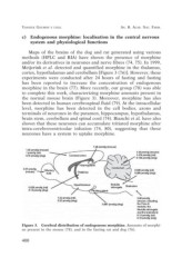
Maps of the brains of the dog and rat generated using various
methods (HPLC and RIA) have shown the presence of morphine
and/or its derivatives in neurones and nerve fibres (74, 75). In 1999,
Meijerink et al. detected and quantified morphine in the thalamus,
cortex, hypothalamus and cerebellum [Figure 3 (76)]. However, these
experiments were conducted after 24 hours of fasting and fasting
has been reported to increase the concentration of endogenous
morphine in the brain (77). More recently, our group (78) was able
to complete this work, characterizing morphine amounts present in
the normal mouse brain (Figure 3). Moreover, morphine has also
been detected in human cerebrospinal fluid (79). At the intracellular
level, morphine has been detected in the cell bodies, axons and
terminals of neurones in the putamen, hippocampus, hypothalamus,
brain stem, cerebellum and spinal cord (74). Bianchi et al. have also
shown that these neurones can accumulate tritiated morphine after
intra-cerebroventricular infusion (74, 80), suggesting that these
neurones have a system to uptake morphine.
Figure 3. Cerebral distribution of endogenous morphine. Amounts of morphi-
ne present in the mouse (78), and in the fasting rat and dog (76).
400