
Page 99 - 73_04
P. 99
VOL. 73 (4), 901-925, 2007 ROLES OF PROTEIN PHOSPHATASE TYPE 1...
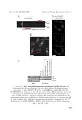
FIGURE 3.- Dis2 internalisation and recruitment to the cell tips. A)
Kymograph of the area between the green lines shows the internalisation
movement of Dis2.NEGFP. B, C) In wsh3? (B) and tea1? (C) cells the
Dis2.NEGFP cap structure at the cell tips was absent. D) Consecutive green and
red images of dis2.NEGFP wsh3.tdTom cells showed the colocalisation of
Dis2.NEGFP and Wsh3.tdTom at cell tips. E) Extracts were prepared and Pk
immunoprecipiates were isolated and blotted with antibodies to recognise the Pk
or GFP epitopes. Dis2 and Wsh3 co-immunoprecipitated. Mutations of the PP1
binding site of Wsh3 (V223A or F225A) abolished Dis2 and Wsh3 interaction.
Bar, 5 µm (B, C, D).
909