
Page 98 - 73_04
P. 98
ISABEL ÁLVAREZ-TABARÉS Y COLS. ANAL. REAL ACAD. NAC. FARM.
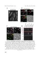
FIGURE 2.- Subcellular localisation of Dis2.NEGFP and Sds21.NEGFP.
A) Dis2.NEGFP localized in the nucleus and diffusely in the cytoplasm. It also
associated with a bright nuclear dot (?), the cell tips (?), cell equator (?) and
endocytic vesicles. B) Sds21.NEGFP localized in the nucleolus, not stained with
Hoescht 33342 which stains the chromatin rich region. C) Sds21.NEGFP in dis2?
cells localized in the nucleolus, the bright nuclear dot and the cell tips (insets).
D) ChIP analysis showed a clear enrichment of the signal arising from PCR with
primers to the central but no other sequences in immunoprecipitates from
dis2.Npk cells (lane 5), but not when antibodies were not conjugated to the
beads (lane 4) or the strain did not contain the tagged dis2+ gene (lane 2). Bar, 5
µm (A, B, C).
908