
Page 114 - 73_02
P. 114
PATRICIA MARÍN-GARCÍA Y COLS. AN. R. ACAD. NAC. FARM.
RESULTS AND DISCUSSION
Previous works have shown that P2X7 receptor activation, and
also reported for P2X4, P2X2, P2X2/P2X3, can lead to open a large
pore through which glutamate can reach the extracellular medium
(5, 6). Since P2X7 is highly expressed in granule neurons where
distributed, mainly, along neuronal fibers (7) we decided to measure
glutamate content in the extracellular media following P2X7 receptor
activation in order to test whether P2X7 receptor could also open
a pore in these cells. Thus, granule neurons growth in culture for
14 days were stimulated with BzATP for 1 minute and the glutamate
content was measured using the Amplex Red Glutamic Acid Assay
Kit (Molecular Probes) according to the protocol provided by the
manufacturer.
The quantification of glutamate using the Amplex® Red Glutamic
Acid/Glutamic Oxidase Assay Kit provides an ultrasentive method for
detecting glutamic levels as low as 10 nM. We also tried to measure
the glutamate release utilising the enzyme-linked fluorometric assay
based on glutamate dehydrogenase activity and the changes in the
fluorescence of NADPH (8), but the glutamate content in the media
was under the limits of detection by this technique (data not shown).
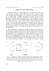
As it can be observed in Figure 1 the principle of the assay is based
FIGURE 1. Principle of coupled enzymatic assays using Amplex Red reagent.
Oxidation of L-Glutamate by glutamate oxidase results in generation of H2O2,
which is coupled to conversion of the Amplex Red reagent to fluorescent resorufin
by HRP. The detection scheme shown here is used in Amplex® Red Glutamic
Acid/Glutamate Oxidase Assay Kit.
446