
Page 91 - 72_04
P. 91
VOL. 72 (4), 629-642, 2006 PROTEIN PROCESSING IN PLASMODIUM FALCIPARUM?
was also designed to use as control. All dsRNAs were obtained from
Dharmacon Research (Lafayette, CO, USA) in annealed and
lyophilised form and were suspended in RNase-Dnase-free water
before use.
Immunodetection
Antibodies were raised against two different recombinant
P. falciparum G6PD-6PDL polypeptides expressed in the vector
pGEX (Amersham Biosciences), which contains the glutathione-
S-transferase sequence upstream from the polylinker to produce
a fusion protein with the insert. Sequences from ntG6PD-6PGL
(AAYYICKEIYDKQQINKDGYVVIGLSGGRTPIDVYKNMCLIKDIKIDKSKL)
and ctG6PD-6PGL (KILKSIPSIKLEDTIIGQYEKAENFKEDENNDDESKKNHS)
(see Figure 1 for their location within the G6PD protein) were
amplified and cloned into pGEX. Expression in E. coli was achieved
following the manufacturer’s instructions and the two glutathione-S-
transferase/G6PD-6PDL fusion proteins were separately purified using
a glutathione sepharose affinity column (Amersham Biosciences).
Cleavage of the fusion protein by factor X and subsequent separation
in SDS-PAGE provided pure protein for antibody production.
Antibodies against ntG6PD-6PGL and ctG6PD-6PGL where raised
separately in rabbits but used as a mixture in the Western blot analyses
to increase the signal.
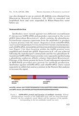
anti-6PGL- anti-G6PD-
domain domain
anti-6PGL domain (AAYYICKEIYDKQQINKDGYVVIGLSGGRTPIDVYKNMCLIKDIKIDKSKL)
anti-G6PD domain (KILKSIPSIKLEDTIIGQYEKAENFKEDENNDDESKKNHS)
FIGURE 1. G6PD-6PGL protein and location of antibody reactivity. Scheme
showing primary structure of the G6PD-6PGL gene with two main functional
domains, the low complexity stretch (black) and the sequences of the peptides
corresponding to the two antibodies raised (see text).
633