
Page 86 - 66_04
P. 86
ARMIN WOLF ANAL REAL ACAD. FARM.
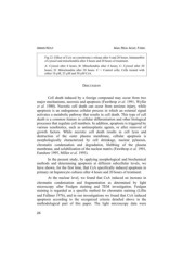
Fig.12: Effect of CsA on cytochrome c release after 4 and 20 hours. Immunoblot
of cytosol and mitochondria after 4 hours and 20 hours of treatment.
A: Cytosol after 4 hours, B: Mitochondria after 4 hours; C: Cytosol after 20
hours; D: Mitochondria after 20 hours. C = Control cells; Cells treated with
either 10 µM, 25 µM and 50 µM CsA.
DISCUSSION
Cell death induced by a foreign compound may occur from two
major mechanisms, necrosis and apoptosis (Fawthrop et al. 1991, Wyllie
et al. 1980). Necrotic cell death can occur from noxious injury, while
apoptosis is an endogenous cellular process in which an external signal
activates a metabolic pathway that results in cell death. This type of cell
death is a common feature in cellular differentiation and other biological
processes that regulate cell numbers. In addition, apoptosis is triggered by
various xenobiotics, such as antineoplastic agents, or after removal of
growth factors. While necrotic cell death results in cell lysis and
destruction of the outer plasma membrane, cellular apoptosis is
morphologically characterized by cell shrinkage, nuclear pyknosis,
chromatin condensation and degradation, blebbing of the plasma
membrane, and solubilization of the nuclear matrix (Fawthrop et al. 1991,
Eanshaw 1995, Miller et al. 1993).
In the present study, by applying morphological and biochemical
methods and determining apoptosis at different subcellular levels, we
have shown, for the first time, that CsA specifically induced apoptosis in
primary rat hepatocyte cultures after 4 hours and 20 hours of treatment.
At the nuclear level, we found that CsA induced an increase in
chromatin condensation and fragmentation as determined by light
microscopy after Feulgen staining and TEM investigation. Feulgen
staining is regarded as a specific method for chromatin staining (Lillie
and Fullmer 1976), and in our investigations we found that CsA induced
apoptosis according to the recognized criteria detailed above in the
methodological part of this paper. The light microscopy data were
24